Common types of hyperpigmentation
Melasma
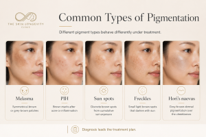
Melasma appears as symmetrical brown to grey brown patches, most often on the cheeks, forehead, upper lip and nose. It is linked to hormones, pregnancy, oral contraceptive use, genetics, heat, visible light and ultraviolet exposure. Melasma is a chronic condition that is managed rather than permanently removed, and treating it too aggressively with lasers can make it worse. Read more in our guide to melasma treatment in Singapore.
Post inflammatory hyperpigmentation
Post inflammatory hyperpigmentation, often shortened to PIH, is the brown staining left behind after inflammation or injury. Acne is the most common trigger in Singapore, but eczema, insect bites, aggressive scrubs and even earlier laser treatments can cause it. PIH is more common and more persistent in darker skin types. Our article on acne marks and PIH treatment in Singapore explains how acne marks differ from true acne scars.
Sun spots and age spots
Sun spots, also called solar lentigines or age spots, are discrete, well defined brown spots on sun exposed areas such as the cheeks, temples and the backs of the hands. They reflect cumulative ultraviolet exposure over the years. Discrete spots like these often respond well to pigment lasers, which is covered in detail in our guide to freckles, sun spots and age spots.
Freckles
Freckles are small, light brown spots that usually appear in childhood or the teenage years and darken with sun exposure. They are partly genetic and partly sun related. Freckles typically lighten well with pigment lasers, although new freckles can appear with ongoing sun exposure if protection is inconsistent.
Hori’s naevus and dermal hyperpigmentation
Hori’s naevus, also known as acquired bilateral naevus of Ota like macules or ABNOM, presents as grey brown to slate coloured macules over both cheekbones. The pigment sits in the dermis, which is deeper than the pigment in melasma or freckles. Dermal pigmentation does not respond to creams and needs a different laser strategy over a longer course. It is also commonly mistaken for melasma, and the two frequently coexist. Our article on stubborn pigmentation in Asian skin covers this in depth.
What recent research tells us about hyperpigmentation
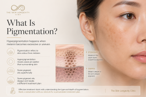
Modern research has reshaped how doctors understand these conditions, and it reinforces the diagnosis first approach to treatments for best results.
Melasma is no longer seen as a pathology involving excess melanin in isolation. It involves melanocytes, surrounding skin cells, the fibroblasts in the deeper skin, mast cells and the small blood vessels of the dermis, with inflammation and increased blood vessel activity both contributing to the pigment. This is why calming and protecting the skin matters as much as targeting the colour, and why aggressive heat based treatment tends to backfire in melasma.
Sun spots tell a similar story from a different angle. The skin over a sun spot is visibly thicker than nearby skin, the deeper layer shows the disorganised elastic tissue of long term sun damage, and the damaged fibroblasts there release signals that keep the overlying skin producing pigment. Increased blood vessel density in the deeper skin has been found in both sun spots and melasma, which is one reason plans that address blood vessel activity as well as pigment can perform better.
The practical takeaway is consistent. Pigmentation is driven from more than one layer and more than one cell type, so the most reliable results come from combining sun protection, topical treatment and correctly chosen procedures rather than relying on any single step.
How to tell the difference between pigment types
The table below summarises the features doctors weigh during an assessment. It is a guide for understanding, not a substitute for an in person diagnosis.
Common types of hyperpigmentation in Singapore
|
Pigment type |
Typical appearance |
Usual depth |
Common triggers |
Tendency to recur |
|
Melasma |
Symmetrical brown or grey brown patches on
cheeks, forehead, upper lip |
Epidermal, dermal or mixed |
Hormones, pregnancy, heat, ultraviolet and
visible light |
High, needs long term control |
|
PIH |
Brown marks at sites of previous acne or
injury |
Epidermal or dermal |
Acne, eczema, trauma, procedures |
Recurs if inflammation continues |
|
Sun spots |
Discrete, well defined brown spots on sun
exposed skin |
Epidermal |
Cumulative sun exposure |
New spots form with ongoing exposure |
|
Freckles |
Small scattered light brown spots from a
young age |
Epidermal |
Genetics plus sun exposure |
Darken and reappear with sun |
|
Hori’s naevus |
Grey brown macules over both cheekbones |
Dermal |
Genetic predisposition in East Asian skin |
Pigment persists without laser treatment |
A thorough clinical history, examination under good lighting and sometimes assessment tools that highlight pigment depth all contribute to the diagnosis. Because several types often coexist on the same face, a treatment plan usually has to address more than one condition at a time. This is one of the reasons why a detailed consultation and evaluation precedes any laser recommendation at our clinic.
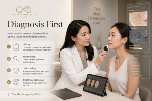
Pigmentation treatment in Singapore should begin with diagnosis, because different pigment types require different timelines, treatments and maintenance plans.
How doctors assess pigmentation before recommending treatment
A pigmentation consultation is a diagnostic exercise before it is anything else. Expect your doctor to work through several layers.
The history comes first: When the pigment appeared, whether it fluctuates, what makes it darker, pregnancy and hormonal history, contraceptive use, medications, previous treatments and their outcomes, your sun exposure patterns and your current skincare all narrow the differential. A patch that appeared in the months after starting a contraceptive pill points differently from spots that have accumulated slowly since your thirties, or marks that map onto last year’s breakouts.
Examination follows, under good lighting and often with magnification. The doctor assesses colour tone, since brown suggests superficial pigment while grey or slate tones suggest dermal pigment, along with the pattern, symmetry, border definition and distribution across the face.
Finally comes lesion safety. Any pigmented spot that is new, changing, irregular in border or colour, or simply different from its neighbours deserves medical scrutiny before any cosmetic treatment. Lasering an undiagnosed lesion is never acceptable practice. Confirmed benign lesions can then be treated on their merits, as described on our
benign skin lesion removal page.
Only after these steps does treatment selection begin, and by then the plan has usually chosen itself, because each pigment type carries its own playbook.

Pico laser may help selected pigmentation types, but the safest treatment plan depends on pigment depth, skin tone and recurrence risk