

31 Aug Pigmentation Returning or Darker After Laser: Why It Happens in Asian Skin
Why pigmentation comes back after laser
By Dr Rachel Ho | Aesthetic Doctor, Founder, The Skin Longevity Clinic, Singapore
Medically reviewed by Dr Rachel Ho, MBBS, MRCS, MMed (ENT), 7 October 2026.
Pigmentation that survives creams, peels and several laser sessions is often being treated as one problem when it is actually two or three. Across the cheeks, melasma, Hori’s naevus, sun spots and post inflammatory hyperpigmentation can overlap, even though they sit at different depths and respond to different treatments.
A stronger laser setting is therefore rarely the first answer. The useful first step is to identify which pigment is present, how reactive the skin is and whether the concern is superficial, dermal or mixed. International laser guidance similarly emphasises that the diagnosis, pigment depth and skin phototype should determine whether laser treatment is appropriate and which treatment parameters are used1.
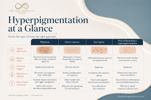
Melasma, Hori’s naevus, sun spots and post inflammatory hyperpigmentation can look similar but behave differently. Pigment depth, triggers and skin reactivity help determine whether skincare, photoprotection, laser treatment or a combination is appropriate.
Pigmentation at a glance
Common types of hyperpigmentation in Singapore at a glance
| Pigmentation condition | Typical appearance | Usual pigment depth | Common triggers or behaviour | Response to skincare | Role of laser treatment |
|---|---|---|---|---|---|
| Melasma | Soft edged brown or grey brown patches, usually appearing symmetrically across the cheeks, forehead, nose or upper lip | Epidermal, dermal or mixed | Often darkens with ultraviolet exposure, visible light, heat and hormonal changes. Recurrence is common | Topical pigment regulators, barrier care and consistent photoprotection can help control the condition | Laser treatment may be used cautiously as an adjunct after the melasma has been stabilised. Aggressive treatment can trigger rebound pigmentation |
| Hori’s naevus | Speckled grey brown, slate or bluish macules over both cheekbones, sometimes extending towards the temples or nose | Dermal | Usually develops during adulthood and tends to remain relatively stable rather than fluctuating with hormones or sunlight | Skincare has very limited effect because the pigment producing cells lie within the dermis | Picosecond or Q switched pigment lasers are commonly used over a staged course. Temporary darkening or post inflammatory hyperpigmentation can occur |
| Sun spots | Discrete, clearly defined brown or dark brown spots on sun exposed areas | Mainly epidermal | Develop gradually through cumulative ultraviolet exposure. New spots may continue to appear with further sun exposure | Skincare may produce modest lightening. Photoprotection helps prevent new spots. | Targeted pigment laser treatment often produces a faster and more predictable response than treatment for melasma |
| Post inflammatory hyperpigmentation | Brown, grey or occasionally blue grey marks appearing after acne, eczema, irritation, picking, burns or procedures | Epidermal, dermal or mixed | Follows inflammation or injury and may persist longer in Asian and more melanised skin | May help selected cases. Excessive inflammation can worsen PIH. | Selected cases may benefit from laser treatment, but excessive heat or inflammation can make the pigmentation darker |
| Mixed pigmentation | A combination of blended patches, grey speckles and clearly defined dark spots within the same area | Multiple depths | Melasma, Hori’s naevus, sun spots and post inflammatory pigmentation may coexist on one face | Skincare may improve the superficial and reactive components while leaving dermal pigment unchanged | Treatment is usually staged. Reactive pigmentation is stabilised first, followed by targeted treatment for the deeper or more defined |
Hyperpigmentation is an umbrella term for areas of skin that appear darker because of increased melanin production or abnormal pigment deposition. Dark spots may look similar in a photograph, but the pattern, depth, trigger and likelihood of recurrence can be very different.
This is why there is no universal answer to the search for pigmentation treatment in Singapore. A treatment that clears an isolated sun spot may irritate melasma, while a cream that improves post acne dark spots will rarely remove Hori’s naevus.
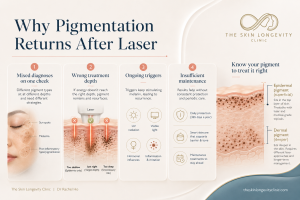
Why do dark spots return after laser? Common explanations include overlapping pigment conditions, treatment at the wrong depth, continuing ultraviolet or hormonal triggers and insufficient maintenance after improvement.
Why does pigmentation keep returning after treatment?
More than one pigment condition may be present
One cheek can contain a blended melasma patch, several sun spots and speckled dermal pigment from Hori’s naevus. Treating only the superficial brown pigment can create some initial improvement while leaving the deeper grey component unchanged.
The patient may understandably conclude that the laser failed. In reality, the treatment may have improved one part of a mixed pigmentation pattern while another component continued to show through.
The treatment may not have matched the pigment depth
Topical pigment regulators work mainly by reducing pigment production, pigment transfer or epidermal cell turnover. They can be valuable for melasma and post inflammatory hyperpigmentation, but they rarely clear the dermal melanocytes responsible for Hori’s naevus.
Superficial chemical peels also have limited influence on deep dermal pigment. Conversely, settings used to target deeper pigment may produce too much inflammation for reactive melasma prone skin1,2.
The biological triggers may still be active
Laser treatment can remove or fragment existing pigment without switching off every process that created it. Ultraviolet exposure, visible light, hormones, inflammation and chronic photodamage can continue to stimulate melasma after a technically successful session.
This distinction is especially relevant in Singapore, where sunlight exposure occurs throughout the year. Maintenance therefore forms part of the treatment rather than something added only after the pigmentation returns3,10.
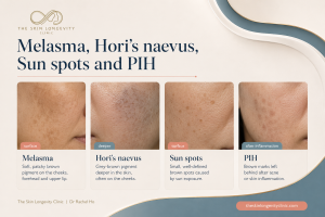
Pattern and colour provide useful clues to the type of pigmentation. Melasma usually forms blended patches, Hori’s naevus appears grey and speckled, sun spots are discrete, while PIH develops after acne, irritation or skin injury.
Which pigment was it?
The next step after a poor laser result depends on what was treated. Symmetrical patches that fluctuate with sun and hormones are usually melasma: see melasma treatment in Singapore. Sharp-edged spots and small light-brown dots are sun spots and freckles: see freckles, sun spots and age spots. Grey-brown speckles over both cheekbones that ignore creams suggest Hori’s naevus, a dermal pigment. Brown marks after acne or irritation are PIH: see acne marks and PIH treatment. Most faces with stubborn pigmentation have more than one of these. The full comparison is in our pigmentation treatment guide.
How can melasma, Hori’s naevus and sun spots be distinguished?
Colour provides an important clue. Melasma usually forms soft blended patches, Hori’s naevus appears more grey or slate and speckled, while sun spots are discrete and sharply defined.
Behaviour also helps. Melasma often fluctuates with sunlight, hormones and time, whereas Hori’s naevus tends to remain more stable and is largely unresponsive to creams. Sun spots usually darken gradually with cumulative ultraviolet exposure.
A doctor will also consider age of onset, previous inflammation, family history, medication, hormonal history and response to previous treatments. Examination under consistent lighting and selected imaging tools may help, but no photograph or machine reading replaces the clinical history.
Any dark spot that is changing in size, shape, colour or surface should be examined before cosmetic laser treatment. Pigment removal should only proceed after a concerning skin lesion has been appropriately excluded.
Pico laser VS Q-switched laser: the differences
| Type of laser | Picosecond laser | Q switched laser |
|---|---|---|
| Indications | Selected epidermal and dermal pigmentation, depending on wavelength and mode | Selected epidermal and dermal pigmentation, with a long clinical history in pigment treatment |
| Main consideration | Shorter pulse duration may increase photoacoustic pigment fragmentation | Established pigment platform with outcomes dependent on wavelength, fluence and technique |
| Clinical takeaway | Diagnosis and treatment parameters determine suitability | Diagnosis and treatment parameters determine suitability |
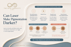
A treated dark spot may temporarily darken before it fades. Persistent, spreading or uneven darkening after laser may represent post inflammatory hyperpigmentation or a melasma flare and should be reviewed before another session.
Why can laser treatment make pigmentation darker?
A laser creates a controlled interaction between energy and pigment. When the surrounding skin receives excessive thermal or inflammatory injury, melanocytes may respond by producing more melanin.
This response can result in post inflammatory hyperpigmentation or a flare of melasma. The risk is particularly relevant in more,4 melanised skin and when treatment settings are too aggressive for the condition being treated1.
Repeated low energy laser toning also carries risk when cumulative exposure becomes excessive. A systematic review found reports of persistent mottled hypopigmentation, inflammatory hyperpigmentation and melasma recurrence after low fluence Q switched Nd:YAG treatment9.
Some discrete dark spots become temporarily darker before the treated pigment sheds or fades. Diffuse darkening, prolonged redness, new blotchy pigmentation or pale speckled areas deserve review rather than another identical session.
PIH after laser: how long it lasts
Post-inflammatory hyperpigmentation (PIH) is the most common reason pigment looks darker a few weeks after laser in Asian skin. Its course is chronic and unpredictable. Brown, epidermal PIH fades faster and responds better to treatment than grey-brown, dermal PIH13.
Pulse type matters. In darker skin, PIH occurred in 22% of patients after Q-switched alexandrite treatment for freckles and sun spots, against 6% after long-pulsed treatment14. The first step is to stop further laser sessions and control any inflammation, then treat the pigment13.
Melasma rebound after laser toning
Melasma can return or darken after a course of laser toning. In a split-face trial, melasma recurred on 12.5% of sides after both picosecond and nanosecond 1064 nm treatment15. A meta-analysis found low-fluence Q-switched Nd:YAG laser alone performed no better than medical treatment, while laser combined with medication gave more improvement16.
Combining toning with fractional microneedle radiofrequency lowered the rate of rebound hyperpigmentation and mottled white spots compared with toning alone in Asian melasma patients17.
White spots after laser toning
Repeated low-fluence Q-switched laser toning can leave pale, speckled spots (mottled or guttate hypopigmentation). In a case series of 14 Chinese women who had 6 to 50 toning sessions, all developed mottled depigmentation, and the 5 treated for melasma saw no improvement in their melasma. Depigmentation can appear after only a few sessions. Targeted narrowband UVB helped repigment the skin in the 5 patients who had it18.
What to do in the first 4 weeks after pigment darkens
- Stop further laser sessions until a doctor has reviewed the skin.
- Use a tinted sunscreen with visible-light protection every day. Visible-light protection improved melasma more than UV-only sunscreen in a randomised trial, and a tinted sunscreen kept melasma skin more even through summer19,20.
- Repair the skin barrier with a gentle cleanser and moisturiser, and stop scrubs, peels and strong acids.
- Start prescribed topical treatment. Triple combination cream cleared or nearly cleared melasma in 77% of patients at 8 weeks21. Oral tranexamic acid may be considered for melasma after screening22.
- Take photographs in the same light each week, so the doctor can tell PIH, melasma flare and hypopigmentation apart before any further treatment.
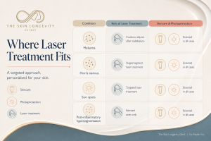
Laser treatment has a different role in each pigmentation condition. Melasma requires cautious treatment after stabilisation, Hori’s naevus usually needs a staged pigment laser course, sun spots may respond to targeted treatment and PIH requires careful patient selection. Skincare and photoprotection remain important throughout.
What is the role of skincare when creams have already failed?
Creams that fail to clear Hori’s naevus may still be useful for the melasma or PIH sitting around it. Skincare can suppress new pigment production, reduce inflammation, support the skin barrier and maintain improvement between laser sessions.
Depending on the diagnosis, medical suitability and skin tolerance, options may include hydroquinone, azelaic acid, retinoids, cysteamine, niacinamide or tranexamic acid. Combining several strong active ingredients without a plan can create irritation and more pigmentation, so the routine should remain manageable4,12.
Photoprotection is central to any melasma or hyperpigmentation plan. In a randomised trial, sunscreen providing ultraviolet and visible light protection improved the response to hydroquinone more than ultraviolet protection alone, supporting the use of suitable visible light protection in melasma10.
Tinted sunscreens containing iron oxides may improve visible light protection, although products differ in tint, coverage and formulation. A high SPF number alone does not describe the full visible light protection of a product.
What can a combined pigmentation treatment plan look like?
Step 1: identify every pigment type
The consultation should map melasma, Hori’s naevus, sun spots and PIH separately. Skin tone, barrier condition, recent tanning, previous reactions and active inflammation should also influence the plan.
Step 2: stabilise reactive pigmentation
Melasma and active PIH usually need photoprotection, gentle barrier care and appropriate topical treatment before significant laser exposure. This stage reduces ongoing pigment stimulation and helps the skin tolerate later procedures.
Step 3: treat the selected pigment target
A defined sun spot may receive targeted treatment, while Hori’s naevus generally needs a staged dermal pigment laser course. Melasma may receive cautious low energy laser only when it is sufficiently stable and the likely benefit outweighs the risk.
Step 4: review the response before escalating
Progress should be assessed using photographs taken under consistent lighting. A treatment that creates excessive redness, PIH or uneven lightening should be reconsidered rather than automatically repeated at a stronger setting.
Step 5: maintain the result
Sunscreen, visible light protection, suitable topical treatment and periodic review help reduce recurrence. Maintenance is especially important for melasma because clearing existing pigment does not remove its underlying tendency to reactivate.
How long does pigmentation laser treatment take?
Clearly defined sun spots may lighten after one or a small number of sessions, although results vary. Hori’s naevus usually clears gradually over a course because the pigment lies within the dermis.
Melasma and PIH often follow a less linear timeline. Improvement may occur over several months, while continued maintenance is needed to reduce rebound and recurrence4,5,7.
A package promising that every form of pigmentation will disappear after the same number of sessions overlooks these differences. The expected course should follow the diagnosis rather than the device being sold.
Dr Rachel Ho’s clinical perspective on stubborn pigmentation in Singapore
I often meet patients who believe their pigmentation is resistant because several lasers or creams have failed. More commonly, the face has been treated as one colour problem when it contains several separate conditions.
My approach to pigmentation in Asian skin begins with identifying what is superficial, what is dermal and what is biologically reactive. A measured sequence usually produces a safer and more sustainable result than repeatedly increasing the laser energy whenever pigment remains.
Frequently asked questions about pigmentation treatment in Singapore
Why do dark spots return after laser treatment?
Pigmentation may return because only one part of a mixed condition was treated, the pigment depth was misjudged or the biological trigger remained active. Melasma is particularly likely to recur when photoprotection and maintenance treatment stop.
Can laser remove pigmentation permanently?
Selected sun spots and Hori’s naevus may achieve substantial, durable improvement, although new sun spots can develop and some dermal pigment may require further treatment. Melasma is usually controlled rather than permanently cured.
How many laser sessions are needed for pigmentation?
The number varies with the diagnosis, depth, treatment wavelength, skin tone and response. A discrete sun spot may need few sessions, while Hori’s naevus or mixed pigmentation generally requires a longer staged course.
How can I tell melasma from Hori’s naevus?
Melasma usually forms blended brown patches that fluctuate with light, hormones and time. Hori’s naevus more often appears as stable grey or slate speckles across both cheekbones and shows little response to creams.
Can Hori’s naevus be treated with creams?
Topical treatment has very limited ability to clear Hori’s naevus because the melanocytes are situated in the dermis. Creams may still help any surrounding melasma, sun damage or post inflammatory pigmentation.
Can Pico laser make pigmentation worse?
Any laser can worsen pigmentation when the diagnosis, wavelength, energy or treatment interval is unsuitable. Picosecond pulse duration may reduce some thermal effects, but it does not eliminate the risks of PIH, melasma flare or hypopigmentation6,8,9.
Why did my pigmentation become darker after laser?
Brief darkening of a treated superficial spot can occur during the clearance process. Persistent or spreading darkening may represent PIH or a melasma flare and should be reviewed before further laser treatment.
Related reading on The Skin Longevity Clinic
For a broader overview of melasma, PIH, freckles, sun spots and treatment options, read Pigmentation Treatment in Singapore for Asian Skin. Readers considering a device based treatment can continue with pico laser in Singapore.
For recurring symmetrical patches, read Melasma Treatment in Singapore: Why It Comes Back and Where Pico Laser Fits. The guide explains why photoprotection and topical control remain the foundation even when laser forms part of the plan.
Readers with isolated brown spots can continue with Freckles, Sun Spots and Age Spots: Which Pigmentation Responds Best to Laser. Those whose dark marks developed after acne or irritation may find PIH Treatment in Singapore and When Pico Laser Helps more relevant.
The Pico Laser Price Singapore guide explains why treatment cost depends on pigment type, depth, area, number of sessions, aftercare and maintenance. A meaningful quotation should follow the diagnosis rather than precede it.
Dark marks that follow acne, irritation or an earlier laser are explained in our guide to post inflammatory hyperpigmentation.
Conclusion on stubborn pigmentation in Singapore
Stubborn pigmentation is rarely solved by choosing the strongest laser or the newest platform. Better results begin by distinguishing melasma, Hori’s naevus, sun spots and PIH, then matching each condition to the appropriate depth and treatment intensity.
Asian skin can respond well to pigmentation treatment when inflammation is controlled and the laser plan respects the skin’s pigment response. Diagnosis, conservative sequencing and maintenance turn laser treatment from a repeated experiment into a coherent medical plan.
Scientific references
- Passeron T, Genedy R, Salah L, et al. Laser treatment of hyperpigmented lesions: position statement of the European Society of Laser in Dermatology. Journal of the European Academy of Dermatology and Venereology. 2019;33:987 to 1005.
- Park JM, Tsao H, Tsao S. Acquired bilateral nevus of Ota like macules, Hori nevus: etiologic and therapeutic considerations. Journal of the American Academy of Dermatology. 2009;61:88 to 93.
- Liu W, Chen Q, Xia Y. New mechanistic insights of melasma. Clinical, Cosmetic and Investigational Dermatology. 2023;16:429 to 442.
- Mar K, Khalid B, Maazi M, et al. Treatment of post inflammatory hyperpigmentation in skin of colour: a systematic review. Journal of Cutaneous Medicine and Surgery. 2024;28:473 to 480.
- Mardani G, Nasiri MJ, Namazi N, Farshchian M, Abdollahimajd F. Treatment of solar lentigines: a systematic review of clinical trials. Journal of Cosmetic Dermatology. 2025;24:e70133.
- Hu S, Yang CS, Chang SL, et al. Efficacy and safety of the picosecond 755 nm alexandrite laser for treatment of dermal pigmentation in Asians: a retrospective study. Lasers in Medical Science. 2020;35:1377 to 1383.
- Cho SB, Park SJ, Kim MJ, Bu TS. Treatment of acquired bilateral nevus of Ota like macules using 1064 nm Q switched Nd:YAG laser with low fluence. International Journal of Dermatology. 2009;48:1308 to 1312.
- Feng J, Shen S, Song X, et al. Efficacy and safety of picosecond laser for the treatment of melasma: a systematic review and meta analysis. Lasers in Medical Science. 2023;38:84.
- Lee YS, Lee YJ, Lee JM, et al. The low fluence Q switched Nd:YAG laser treatment for melasma: a systematic review. Medicina. 2022;58:936.
- Castanedo Cazares JP, Hernandez Blanco D, Carlos Ortega B, Fuentes Ahumada C, Torres Alvarez B. Near visible light and ultraviolet photoprotection in the treatment of melasma: a double blind randomised trial. Photodermatology, Photoimmunology and Photomedicine. 2014;30:35 to 42.
- Mar K, Khalid B, Maazi M, et al. Prevention of post inflammatory hyperpigmentation in skin of colour: a systematic review. Australasian Journal of Dermatology. 2025.
- Gan C, Rodrigues M. An update on new and existing treatments for the management of melasma. American Journal of Clinical Dermatology. 2024;25:717 to 733.
- Chaowattanapanit S, et al. Postinflammatory hyperpigmentation: A comprehensive overview: Treatment options and prevention. J Am Acad Dermatol. 2017;77(4):607–621. doi:10.1016/j.jaad.2017.01.036.
- Ho SG, et al. A comparison of Q-switched and long-pulsed alexandrite laser for the treatment of freckles and lentigines in oriental patients. Lasers Surg Med. 2011;43(2):108–113. doi:10.1002/lsm.21045.
- Feng J, et al. Comparison of Picosecond and Nanosecond Nd:YAG 1064-nm Lasers in the Treatment of Melasma: A Split-Face Randomized Clinical Trial. Plast Reconstr Surg. 2023;151(4):772–777. doi:10.1097/PRS.0000000000009994.
- Chen J, et al. Efficacy of low-fluence 1064 nm Q-switched Nd:YAG laser for the treatment of melasma: a meta-analysis and systematic review. J Cosmet Dermatol. 2022;21(7). doi:10.1111/jocd.15126.
- Kwon HH, et al. Combined treatment of melasma involving low-fluence Q-switched Nd:YAG laser and fractional microneedling radiofrequency. J Dermatolog Treat. 2019;30(4):352–356. doi:10.1080/09546634.2018.1516858.
- Chan NP, et al. A case series of facial depigmentation associated with low fluence Q-switched 1,064 nm Nd:YAG laser for skin rejuvenation and melasma. Lasers Surg Med. 2010;42(8):712–719. doi:10.1002/lsm.20956.
- Castanedo-Cazares JP, et al. Near-visible light and UV photoprotection in the treatment of melasma: a double-blind randomized trial. Photodermatol Photoimmunol Photomed. 2014;30(1):35–42. doi:10.1111/phpp.12086.
- Polena H, et al. Comparison of Visible Light-Protective Tinted Sunscreen to Untinted Sunscreen to Protect Melasma Patients During Summer: A Prospective Randomized Investigator-Blinded Study. J Cosmet Dermatol. 2025;24(10):e70450. doi:10.1111/jocd.70450.
- Torok HM. A comprehensive review of the long-term and short-term treatment of melasma with a triple combination cream. Am J Clin Dermatol. 2006;7(4):223–230. doi:10.2165/00128071-200607040-00003.
- Del Rosario E, et al. Randomized, placebo-controlled, double-blind study of oral tranexamic acid in the treatment of moderate-to-severe melasma. J Am Acad Dermatol. 2018;78(2):363–369. doi:10.1016/j.jaad.2017.09.053.
- Wong THS, et al. Picosecond Laser Treatment for Acquired Bilateral Nevus of Ota-like Macules. JAMA Dermatol. 2018;154(10):1226–1228. doi:10.1001/jamadermatol.2018.2671.